Search This Blog

CLINICAL BLOOD HAEMATOLOGY - ERYTHROCYTE SEDIMENTATION RATE (ESR) - VSH

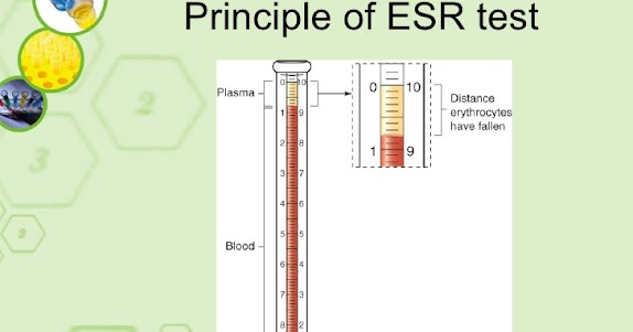
 ESR: the rate of red blood cell/erythrocytes sedimentation/aggregation. The new version of this examination is the "VSH": the Velocity of the Sedimentation of the red blood cells/Haematias. 


Erythrocyte sedimentation is the aggregation of red blood cells in the form of a column. The rate of erythrocyte sedimentation (Erythrocyte sedimentation rate ESR): the measuring of red blood cell aggregation. 


In certain conditions (that cause the growth of acute phase proteins or immunoglobulins), plasma proteins attach to the red blood cell surface and reduce the surface potential, causing red blood cell aggregation and increased sedimentation. 

ESR: the rate at which red blood cells aggregate/settle in an anticoagulated blood sample within an hour. 

Normal ESR range: 0-22mm/h for men and 0-29mm/h for women.


CLINICAL BLOOD HAEMOSTASIS - COAGULATION

COAGULATION: FIBRINOGEN, PT, BT, TT, APTT.

FIBRINOGEN

Fibrinogen/Coagulation Factor I is a plasma glycoprotein produced by the liver. Fibrinogen is the action substrate for both thrombin (final coagulation enzyme) and plasmin (fibrinolytic system enzyme). Fibrinogen belongs to the acute phase proteins (appearing at 24-48 hours post-event). 

Defective fibrinogen levels show liver disfunction. Fibrinogen levels help assessing the body's ability to form a blood clot.  

While coagulation approaches its end, soluble fibrinogen is transformed into insoluble fibrinogen fibres. Fibrinogen fibres protect the wound by intercalating; along with the platelets, they form a clotted blood barrier that blocks bleeding until healing. 

Fibrinogen analysis measures the soluble amount of Factor I (fibrinogen dissolved in the blood), before its conversion into insoluble fibrin.

Fibrinogen normal levels: 200-400 mg/dL (according to the laboratory reference values - reactive used etc.).
 
 
PROTHROMBINE TIME (PT; INR): 

Prothrombin time (PT) evaluates the activity of factors involved in the extrinsic and common coagulation pathway. Evaluates both the activity of vitamin K-dependent coagulation factors (except F IX), factor V and fibrinogen, as well as the function of liver protein synthesis, with diagnostic and therapeutic implications. 

In vitro, main route of blood coagulation initiation: the extrinsic system. This includes blood components and vascular elements, the initiation of coagulation occurring when the tissue factor (FT) is bound to F VIIa. 
The F VIIa - FT enzyme complex activates both F IX and F X. 
F Xa interacts with its cofactor F Va, forming the prothrombinase complex; the complex: enough to generate few thrombin amounts next to FT-expressing cells. 

In vitro, PT detects clot formation. This represents fibrin polymerization, resulting from thrombin action.
Being absent in normal plasma, the tissue factor (FT) must be provided from an external source. This is why the cascade of enzymatic reactions triggered in PT is known as the "extrinsic pathway".  

Prolonged PT indicates deficiency of coagulation factors (I, II, V, VII, X) or an inhibitor's presence. 

PT is the most commonly used test monitoring oral anticoagulant therapy. Results may be expressed as follows:
- as clotting time - in seconds; PT - time (s); normal: 11-13.5 s;
- as a percentage (%) of normal prothrombin activity; measurable range: 12.5-120%; AP - volume fraction (%)
- as prothrombin ratio (PR = PT patient in seconds / normal plasma PT in seconds); PR - prothrombin time ratio (1)
- as INR - International Normalized Ratio - (1); normal: 0.8-1.1.
INR = (PT patient / PT normal plasma) x ISI; ISI: international sensitivity index of used thromboplastin, calculated in report to reference thromboplastin for which ISI = 1. 

INR = 2.0-3.0 target of oral anticoagulant treatment for most clinical cases;
INR = 2.5-3.5 target in anticoagulant treatment for: recurrent deep vein thrombosis, recurrent systemic embolism, cardiac stent, mechanical heart valves.


BLEEDING TIME (BT/CT)

A test that investigates primary haemostasis, thus being an indicator of the vascular and platelet phases efficiencies. 

BT depends on the:
- platelets (function and number)
- adhesion plasma proteins
- vascular wall matrix integrity
Critical values:  >15 minutes. 

Bleeding Time is increased when platelet levels are low or when platelets are qualitatively abnormal. 


THROMBIN TIME (TT)

TT measures the time of fibrin formation under the action of thrombin, and its aggregation to form insoluble clot - the final coagulation step. Assesses the activity of fibrinogen.

Under thrombin action on fibrinogen, fibrinopeptides A and B are released. Upon their cleavage, fibrin monomers form soluble aggregates. 
Due to the action of Factor XIII (activated by thrombin), along with that of Ca ions, a transverse polymerization of fibrin monomers takes place, with the formation of insoluble fibrin. 


Thrombin Time mainly reflects the function and interaction between the exogenous thrombin and the endogenous fibrinogen. 

TT measurable range: 13-240 seconds. Thrombin clotting time is generally <22s, between 14-16s (every lab sets its own reference values, according to the reagent kits used; other labs may have: 7,0-12,0s). Critical values: >60s. 


ACTIVATED PARTIAL THROMBOPLASTINE TIME (APTT)

Also called Partial Thromboplastin Time (PTT), is a functional test evaluating both "intrinsic" and "common" coagulation pathways. 
In vitro, the contact system is involved when blood interacts with a foreign surface, such as the cardiopulmonary bypass. 

Kallikrein cleaves HK, releasing bradykinin and kinin-free kininogen (activated HK). 
In vitro, activation of HK is achieved by adding:
- "partial thromboplastin"; composed only of phospholipids; no protein, no TF;
- silica - a surface activator; provides the negatively charged surface.
Than, calcium chloride is added and time expressed in seconds is measured until the clot's formation. 
The so-called "partial thromboplastin" does not contain the tissue factor (protein) needed to initiate coagulation in PT, as in "complete thromboplastin" stage. The silica provides the negatively-charged surface needed for coagulation contact pathway activation. 

The cascade of reactions triggered in PTT were called as "intrinsic pathway" based on the misconception that coagulation is initiated without the addition of any external factors. The external factor involved in initiating coagulation in PTT: the negatively-charged glass surface of the reaction tube; this can be potentiated by adding silica, kaolin, ellagic acid, in what we now refer to as the "activated" partial thromboplastin test.  

Deficiency/inhibition of HK, prekalikrein or Factors XII, XI, IX and VIII causes prolongation of aPTT with normal PT, while deficiencies of "common pathway" coagulation factors (X, V, II, Fibrinogen) can prolong both aPTT and PT. aPTT is not influenced by FVII or FXIII deficiencies. 

Normal aPTT: 21-35s (therapeutic: 2,0-2,5x normal)


COAGULATION: 


"INTRINSIC PATHWAY": prekalikrein, high-molecular weight kininogen-HK, Factors XII, XI, IX and VIII;
"COMMON PATHWAY": Factors X, V, II and I. 


CLINICAL BLOOD HAEMOSTASIS - BLOOD TYPE IDENTIFICATION

 BLOOD TYPE IDENTIFICATION: Based on characterization of an individual's blood according to the presence/absence of an antigen on the surface of his erythrocytes. Most blood group antigens are glycoprotein in nature and genetically stable. 

The antigen-antibody reaction in which the blood group antigens and their specific antibodies participate is one of agglutination (of red blood cells). Hence, the antigens are also called agglutinogens and the antibodies also agglutinins.  

In current medical practice, the ABO and Rh systems are important. 

The main blood groups: 

A

B

AB

0.

Rh factor (Rhesus) is a group of antigens that may be present or absent on the surface of erythrocytes. Most people have Rh factor present on erythrocytes, so they are called Rh positive. Rh negative: do not have the Rh factor present. 

Incompatibility of Rh positive blood with Rh negative blood is an important cause of transfusion reactions and haemolytic diseases in newborns. 


                                BLOOD TYPE COMPATIBILITY RESUME

                                            +   : Rh present;     -   : Rh absent


MICROORGANISMS IDENTIFICATION - culture growth etc

Microscopy: particularly useful for bacteria identification.

Blood analysis (serology, blood culture etc): identification of microorganisms in blood.

Pharyngeal/nasal exudate, sputum, ocular/otic secretions: collection of biological samples for microbiological examination. 

Urinalysis: first morning urine, from the intermediate jet, after local toilet. Important tool for microorganism presence identification.

Coproparasitological examination, coproculture, from faeces.

Vaginal, urethral, vulvo-vaginal secretion examinations (exudates etc)

Various smears with specific colourings - microscopical examinations of morphology.  

Various molecular genetics techniques for virus identification: RT-PCR, ELISA, Western-Blot, immunological techniques (immunochromatography, immunodifusion, immunofluorescence) etc. 


LABORATORY DIAGNOSIS FOR VIRUSES:


- virus isolation on cell cultures

- viral genome highlighting by gene amplification reaction after reverse transcription (Real-Time Polymerase Chain Reaction - RT-PCR) - frequently used due to high precision

- serological identification - immunochromatography techniques (rapid tests), ELISA, Western Blot etc.


CULTIVATION OF VIRUSES


Viruses have obligatory intracellular parasitism, hence they do not grow on artificial environment/mediums. They can only be cultivated on live substrates:

- laboratory animals - limited use nowadays due to spreading of cell culture usage popularity;

- embryonated chicken egg - offer embryonic tissue for viruses culturing; important in vaccine preparation;

- cell cultures - the mostly used virus-host system in virology research. 

Example: Poliomyelitis

                 - indicated collection of pharyngeal exudate, blood, faeces, cerebrospinal fluid;

                 - isolation of virus on cell cultures; RT-PCR.


CULTIVATION OF BACTERIA ON MEDIA CULTURE:


Microscopical examination: first step to bacteria identification. Main elements to establish bacteria identity: 

- cilia

- capsule

- endospores. 


Morphological examination: on microscopic preparations, fixed and coloured: smears

Smears: obtained by spreading a bacterial colony on a clean and degreased blade, which will be dried, fixed and coloured, for examination under a microscope. 


Culture media - provides nutrients and physico-chemical conditions for bacteria growth;

Sowing - contact of pathological product with the culture medium;

Isolation - single colony transition to other culture medium => pure culture.


MEDIUMS: 


Physical classification:

- solid - reflecting colour, diameter, appearance, media adherence etc;

- liquid - turbidity and turbidity type, surface/bottom film/deposit formation.  

Examples: homogeneous turbidity - S. aureus; surface film - Vibrio cholerae, bottom deposit - Streptococcus pyogenes. 


In terms of composition, media can be:

- simple - simple agar;

- composed - besides the basics, also organic substances (serum, blood) required for special growth care; example: blood agar;

- special mediums - isolation, enrichment, differential. 


BACTERIA CLASSIFICATION:


1. Tinctorial affinity to Gram staining (most important staining, dividing bacteria in Gram-positives - lilac and Gram-negatives - red): 

a) Gram-positives: Streptococcus pneumoniae, Staphylococcus aureus;

b) Gram-negatives: Neisseria gonorrhoeae and meningitidis, Escherichia coli, Helicobacter pylori, Haemophilus influenzae. The Enterobacteriacee family - large number of Gram-negative bacillus species living in the intestines; the most important genre it includes are: Escherichia, Shigella, Salmonella, Klebsiella, Proteus. Enterobacteria classification according to the fermentation capacity of lactose: 

- Pathogenic Enterobacteria: lactose-negative: Salmonella, Shigella;

- Conditioned-pathogenic or non-pathogenic enterobacteria: 

                            a) lactose-positive: Escherichia, Klebsiella

                            b) lactose-negative: Proteus.


2. Need for oxygen:

a) aerobic bacteria: Staphylococcus aureus;

b) anaerobic bacteria: Clostridium botulinum.


3. Glucose fermentation capacity:

a) glucose-fermentative bacteria: Vibrio cholerae;

b) glucose-nonfermentative bacteria: Pseudomonas aeruginosa. 


4. Pathogenicity classification: 

a) nonpathogen bacteria: suboptimal conditions inside human host

b) pathogenic bacteria: always causing illness; example: Treponema pallidum. 

c) conditionate pathogenic bacteria: from normal flora, causing illness in various conditions.  


EXAMPLES OF AGARS AND COLORATIONS FREQUENTLY USED IN MICROBIOLOGY FOR BACTERIA IDENTIFICATION:

- MacConkey Agar: selective and differential culture medium for bacteria. Isolates Gram-negative and enteric bacteria. Differentiates them based on lactose fermentation. Lactose fermenters turn red or pink, non-fermenters do not change colour.

- Kligler's Iron Agar: Enterobacteriaceae identification, based on double sugar fermentation and hydrogen sulphide production.

- Ziehl-Neelsen stain: acid-fast stain identifying acid-fast organisms, mainly Mycobacteria (Nocardia as well). Acid-fast bacilli are bright red after staining. 

- Voges-Proskauer test: detects acetoin in a bacterial broth culture. Red: positive, yellow-brown: negative. Principle: digestion of glucose to acetylmethylcarbinol. 

- BD CLED Agar: CLED: Cystine-Lactose-Electrolyte-Deficient; differential culture medium for bacteria in urine; supports pathogen growth but prevents Proteus species accumulation due to the lack of electrolytes. 


DIAGNOSIS OF PARASITES:


- Coproparasitology: - faeces examination for parasites;

- ELISA;

- Direct agglutinations to detect IgM antibodies;

- Blood culture; cerebrospinal fluid examination for parasites;

- Smear and thick drop examination of peripheral blood; May-Grumwald-Giemsa staining;

- Immunological examination: ELISA, hemagglutination; latex agglutinations

- Parasites/parasite eggs identification in faeces and other material through microscopical examination.

- Macroscopical examination of faeces for parasite and/or parasite eggs elimination;


MICOLOGICAL DIAGNOSIS:


- based on the clinical lesion's aspect and microscopic analysis results of hairs and squamous material collected from lesions.