Search This Blog

GIARDIA LAMBLIA TEST - detection from stool

Giardia Lamblia Antigen


Parasitosis is still a serious global health problem nowadays. 

Giardia Lamblia is the most common protozoan responsible for a wide range of conditions, ranging from severe diarrhea, especially in immunocompromised patients. 

Attachment of the parasite to the duodenal mucosa leads to malabsorption. Intestinal villi atrophy and disappear, leading to significant changes in digestive function, with weight loss and dehydration. In most cases the infection is asymptomatic. 


The Giardia Lamblia quick test on the cassette is a rapid test for immunochromatographic identification and qualitative detection of Giardia Lamblia antigens - 65 kDa coproantigen - a glycoprotein present in Giardia Lamblia cysts and trophozoites. 

Below is presented the work procedure available for most of the quick tests using the immunochromatographic qualitative method of detection.  


IONOMETRY

ION: atom/group of atoms with electric charge (positive charge: cations; negative charge: anions). Many normal substances exist in the body as ions (sodium, potassium, calcium, chloride, bicarbonate, substances known as electrolytes). Chemically, electrolytes are substances that become ions in solution and acquire the capacity to conduct electricity (ions enable the flow of electrical signals through the body). 

The balance of the electrolytes in our bodies is essential for normal function of our cells and organs. If electrolyte levels are too low or too high, cell and organ functions will decline, which could lead to life-threatening conditions. Virtually every metabolic process depends on the presence of electrolytes, which create an "electric potential" needed to do "cellular work".  


The body has complex systems for monitoring and maintaining electrolyte concentrations in a normal narrow range. Physiological functions: 

- maintenance of osmotic pressure and water distribution (water homeostasis)

- conduction of neuromuscular impulses

- acid-base maintenance

- enzyme activation

- electron transfer

- rebuild damaged tissue


Common electrolytes that are measured by doctors with blood testing include sodium, potassium, chloride, bicarbonate, calcium, magnesium, phosphate. These have essential functions in the body. For example, a muscle needs calcium, sodium and potassium to contract. When these substances become imbalanced, it can lead to either muscle weakness or excessive contraction. The heart, muscle and nerve cells use electrolytes to carry electrical impulses to other cells.

Sodium, chloride, potassium, calcium and magnesium (the main electrolytes; nutritional elements) are minerals, and when minerals dissolve in water, they separate into positive and negative ions. For example, when sodium chloride (NaCl) is dissolved in water, it separates into positive sodium ions and negative chloride ions.

Anions: negatively charged ions; migrate towards the anode (positive electrode).

Cations: positively charged ions; migrate towards the cathode (negative electrode).


Major electrolytes (Na+, K+, Cl-, HCO3-) exist as free ions in solution, capable of carrying an electrical charge. Often classified as: major intracellular anion/cation, major extracellular anion/cation. 

Much of the metabolic energy produced by the body is used to establish high intracellular concentrations of potassium (K+) and low concentrations of sodium (Na+), the reverse of the relative concentrations of these ions in the extracellular fluids. The extrusion of sodium requires its movement against a gradient of concentration (higher outside than inside) and electrical potential (inside about 70 mV more negative than outside); work is therefore needed to overcome this electrochemical gradient. 


Functions of the main electrolytes (ions):


- sodium ions: regulate osmotic pressure and the body's water content, transmit nerve signals, contract muscles etc;

- potassium ions: transmit nerve signals and contract muscles including the heart etc;  

- magnesium ions: contract muscles, form bones and teeth, activate enzymes etc;

- calcium ions: transmit nerve signals, contract muscles, form bones and teeth, clot blood;

- chloride ions: regulate osmotic pressure and the body's water content, enable the secretion of stomach acid etc. 

Values differ slightly between serum fluid and plasma fluid; difference is most significant with potassium (serum K+ > plasma K+).

An ion-selective analyser comprises of ion-selective electrode (ISE) and is an analytical technique used to determine the activity of ions in aqueous solution by measuring the electrical potential. It has application in the field of pharmaceuticals and biotechnology. There are four types of ISE: glass electrode, crystalline electrodes, ion-exchange electrodes and enzyme electrodes.


Sodium and potassium and the main and most basic ions analysed. Therefore, more emphasis will be placed on them.  


SODIUM:


Sodium is the major positive ion (cation) in fluid outside of cells. Many processes in the body, especially in the brain, nervous system and muscles require electrical signals for communication. The movement of sodium is critical in the generation of these electrical signals. 


Regulation of osmolality: 

- aldosterone: stimulates sodium (and H2O) retention by kidney (at expense of K+).


Sodium (Na+):

- major cation in ECF (plasma);

- responsible for almost 1/2 the osmotic strength of plasma.


Sodium (Na+) Regulation:

- kidneys are the primary regulators of body sodium and water;

- sodium is freely filtered by glomerulus, 70-80% actively reabsorbed by PCT;

- in times of deficit all sodium is reabsorbed by kidney. 


POTASSIUM:


Potassium is the major positive ion (cation) found inside of cells. Among the many functions of potassium in the body are regulation of the heartbeat and the function of the muscles. 

Potassium levels often change with sodium levels. When sodium levels go up, potassium levels go down, and when sodium levels go down, potassium levels go up. Potassium levels are also affected by a hormone called aldosterone, which is made by the adrenal glands.   

Potassium levels can be affected by how the kidneys are working, the blood pH, the amount of potassium you eat, the hormone levels in your body, severe vomiting, and taking certain medicines, such as diuretics and potassium supplements. Certain cancer treatments that destroy cancer cells can also make potassium levels high. 


Potassium (K+):

- major intracellular cation;

- neuromuscular excitability, heart contractions, maintain ICF volume, maintain H+ concentrations;

- 23x higher in RBC compared to plasma; higher in tissue cells.


Potassium (K+) Regulation:

- Na+-K+-ATP-ase pump continually transports K+ into cells against concentration gradient;

- high intracellular "stores" aid to maintain near-normal extracellular K+ levels in times of deficit;

 - kidney response to conserve K+ is not as immediate and thorough as its response to conserve Na+;

- aldosterone: when sodium is retained, K+ (or H+) is excreted into urine.


CHLORIDE:


Chloride is the major anion (negatively charged ion) found in the fluid outside of cells and in the blood. Chloride plays a role in helping the body maintain a normal balance of fluids. 


Chloride (Cl-):

- major anion in ECF;

- most often Cl- ions shift with Na+ (passive association) and HCO3- to maintain electrical neutrality, osmolality and blood volume. 


Chloride (Cl-) Regulation:

- filtered by glomerulus, passively reabsorbed by the proximal tubules and actively reabsorbed by the ascending Loop of Henle;

- chloride shift: maintains anion-cation balance during buffering process in response to cellular metabolism.


BICARBONATE:


The bicarbonate ion acts as a buffer to maintain the normal levels of acidity (pH) in blood and other fluids in the body. Bicarbonate levels are measured to monitor the acidity of the blood and body fluids.  


Bicarbonate (HCO3-):

- 2nd most abundant anion in ECF;

- maintenance of acid-base balance as the major buffer ion in the carbonic acid/bicarbonate buffer system; major component (>95%) of total CO2 (tCO2).


Bicarbonate (HCO3-) Regulation:

- filtered by the glomerulus and reabsorbed in the proximal and distal tubules;

- kidney has the capacity to reabsorb all or none of the filtered HCO3- as needed to maintain acid/base balance in body.

SODIUM

Sodium: serum and urinary


Serum sodium

All the movements of sodium produce the movement of a variable amount of water. The volume of fluid in the extracellular compartment is directly dependent on the total amount of sodium in the body. The plasma sodium concentration is identical to that of the interstitial fluid. 

In its movements, to achieve an electrical balance, Na+ is followed by anions and primarily by Cl- and HCO3-. Being an alkaline metal and in its movement entraining the bicarbonate anion, Na+ intervenes in maintaining the acid-base balance. Also, Na+ intervenes in the neuromuscular excitability and in the dynamics of the polarization and depolarization phenomena of the cell membrane, opposing the effects of potassium. 

The mechanisms by which the body maintains constant sodium levels in the plasma and extracellular sector are represented by: renal blood flow carbonic anhydrase activity, renin-angiotensin-aldosterone system, ADH, vasopressin, other steroid hormones whose plasma concentration is controlled by he anterior pituitary. 

Biological reference range: premature babies: 132-140 mEq/L; newborns: 133-142 mEq/L; children (1-16): 136-145 mEq/L; adults: 136-145 mEq/L. 


Urinary sodium

The main way to eliminate sodium is through the kidneys. Sodium excretion by the kidneys is influenced by changes in glomerular filtration rate, serum sodium concentration, adrenal cortex activity, the amount of non-resorbable solvates in the filtrate and the volume of extracellular fluid. 

Biological reference range: children: 41-115 mEq/24h; adults: 40-220 mEq/24h.

POTASSIUM

Potassium: serum and urinary.


Serum potassium

Potassium is the main electrolyte (cation) and constituent of the intracellular fluid buffer system. 90% of the potassium is concentrated inside the cell, only small amounts being present in the bones and blood. The damaged cells release potassium into the blood. The entire amount of potassium contained in ingested food is absorbed in the small intestine. The vast majority of potassium (90%) is in ionic form, the rest being protein-related. 

Potassium is indispensable for the normal development of membrane electrical phenomena. It also plays an important role in nerve conduction, muscle contraction, acid-base balance, osmotic pressure, protein anabolism and glycogen formation. Anabolic processes are accompanied by the fixation of potassium in the cell, and catabolic processes by its release. 

Along with calcium and magnesium, potassium controls heart contraction and flow. Potassium and sodium ions are important in the renal regulation of acid-base balance, hydrogen ions being replaced by sodium and potassium ions in the renal tube. Potassium bicarbonate is the major intracellular inorganic buffer. 

Biological reference range:

Premature - umbilical cord blood: 5.0-10.2 mEq/L; premature 48h - venous blood: 3.0-6.0 mEq/L; newborns - umbilical cord blood: 5.6-12.0 mEq/L; newborns - venous blood: 3.7-5.9 mEq/L; children - venous blood: 3.4-4.7 mEq/L; adults: 3.5-5.1 mEq/L. 


Urinary potassium

The greatest amount of potassium is eliminated from the body through the kidneys. Normally 80-90% of potassium is excreted in the urine and the rest in sweat and stool. At the level of the nephron, potassium is subjected to the three fundamental mechanisms: glomerular filtration, complete reabsorption in the proximal tubules and secretion in the distal tubules. 

Biological reference range:

Girls (6-9y): 17-54 mEq/24h; boys (6-9y): 8-37 mEq/24h; girls (10-14y): 22-57 mEq/24h; boys (10-14y): 18-58 mEq/24h; adults: 25-125 mEq/24h.

PHOSPHOROUS

Phosphorous: serum and urinary.


Serum phosphorous

Phosphorous is, after calcium, the most abundant mineral element in the body, being in any tissue. Phosphate is the major intracellular anion. In the cell, phosphorous is mainly involved as organic phosphorous in carbohydrate and lipid metabolism or is bound to proteins and only a small part is present as a phosphate ion. The kidneys are the main regulators of phosphorous homeostasis; approximately 80% of the glomerular filtered amount is reabsorbed in the proximal tubules and 10% in the distal tubules. 

Biological reference range:

Newborns: 4.5-9.0 mg/dL; children: 4.5-5.5 mg/dL; adults: 2.7-4.5 mg/dL. 


Urinary phosphorous

It is recommended to determine urinary phosphorous for the evaluation of phospho-calcium balance and nephrolithiasis. 

Biological reference range: 400-1300 mg/24h; first morning urine: 40-140 mg/dL. 


MAGNESIUM

Magnesium: serum and urinary.


Serum magnesium

Magnesium is an element that, although found in small proportions in the body (0.05% of total body weight), is of great structural and functional importance. About 1% is in plasma, 25% is protein bound, the rest remains in ionized Mg2+ form. In erythrocytes the amount of magnesium is appreciable, about 5.2 mEq/L. Together with Na+, K+ and Ca2+ ions, magnesium regulates neuromuscular excitability and the coagulation mechanisms. The actions of calcium and magnesium are closely linked, the deficiency of one of these elements significantly influencing the metabolism of the other. 

Biological reference range:

Newborns (2 - 4 days old): 1.5-2.2 mg/dL; children (5 months - 6 years old): 1.7-2.3 mg/dL; children (6 - 12 years old): 1.7-2.1 mg/dL; adults: 1.6-2.6 mg/dL. 


Urinary magnesium

Normally 95% of the amount of magnesium that is filtered at the glomerular level is reabsorbed tubularly, especially in the ascending portion of the Henle loop. Magnesium excretion controls the serum level of this element and is dependent on diet. Magnesium, along with calcium, is subject to the effects of parathyroid hormone.  

CALCIUM

Calcium: serum - total and ionic, urinary.


Total serum calcium

Calcium is one of the most important minerals in the body. It is essential for the proper functioning of muscles, nerves, heart, and has an important role in coagulation and bone mineralization. Calcium is the major mineral component of bones. 99% of the body's calcium is in the bones and teeth, which is a huge reservoir for maintaining serum calcium levels, and the rest is distributed in biological fluids and soft tissues. Calcium homeostasis is maintained by parathyroid hormone (PTH). 

About half of the total amount of plasma calcium (45%) is bound to albumin (and only a small portion bound to globulins) in a non-ionized and non-diffusible form, making it a physiological inactive form. A small amount of calcium (5%) is diffusible but not ionized, being represented by citrate, phosphate and calcium bicarbonate. The rest of the plasma calcium is found in ionic or free form and constitutes the physiologically active fraction in the processes of haemostasis and regulation of neuromuscular excitability; its plasma concentration is directly related to PTH and 1,25(OH)2D. 

Calcium in the blood is tested to diagnose and monitor certain diseases related to bones, heart, kidneys and teeth. Its levels do not directly show how much calcium is in the blood, but how much calcium circulates in the blood. Calcium testing can be used if the patient has: kidney stones, bone diseases, neurological disorders.

Abnormal serum calcium levels may indicate parathyroid disfunction, bone disease, carcinoma, malnutrition and malabsorption syndrome, vitamin D deficiency, and kidney disease. Calcium ions play an important role in the transmission of nerve impulses, muscle contraction, heart function and coagulation processes. 

Biological reference range:

0 - 10 days old: 7.6-10.4 mg/dL; 10 days - 3 years old: 6.7-9.8 mg/dL; 3 - 9 years old: 8.4-10.2 mg/dL; 4 - 11 years old: 8.9-10.1 mg/dL; 11 - 13 years old: 8.8 - 10.6 mg/dL; 13 - 15 years old: 9.2-10.7 mg/dL; 15 - 18 years old: 8.4-10.7 mg/dL; adults: 8.8-10.4 mg/dL. 


Serum ionic calcium

Determination of ionic calcium provides guidance on the effect of total protein and albumin on serum calcium levels. A patient may have a high level of total calcium with a normal level of ionic calcium due to an increase in total protein and/or albumin, as is the case with dehydration or multiple myeloma. 

Ionic calcium testing is performed on patients who have a disturbed balance between bound and free calcium, and if they have transfusions, have had major surgery, or have abnormal blood protein levels, such as albumin. High fluctuations in ionic calcium can slow the heart or increase its heart rate, as well as cause muscle spasms, confusion or coma. In the case of very sick patients, it is extremely important to know the level of ionized calcium in order to intervene and prevent major complications. 

Biological reference range:

Newborns: 4.40-5.48 mg/dL; 1-18 years old: 4.80-5.52 mg/dL; adults: 4.65-5.28 mg/dL. 


Urinary calcium

Testing for calcium in the urine shows if the kidneys are excreting the right amount of calcium, and testing for vitamin, phosphorous, and/or magnesium determines what other deficiencies or excesses there are. 

Most of the calcium is excreted in the faeces and a small amount of calcium is excreted in the urine, depending on the calcium intake in the diet. Determination of urinary calcium is important in the diagnosis of hypercalcemia responsible for kidney stones. 

Biological reference range:

Normal diet: 100-300 mg/24h; low calcium diet: 50-150 mg/24h. 


CLINICAL BLOOD HAEMATOLOGY - COMPLETE BLOOD COUNT/CBC

 COMPLETE BLOOD COUNT (CBC) or FULL BLOOD COUNT (FBC), also known as HEMOGRAM/HAEMOLEUCOGRAM: red blood cells (RBCs), white blood cells (WBCs) and platelets (PLTs), concentration of haemoglobin and haematocrit (the volume percentage of red blood cells). HEMOGRAM/HEMOLEUCOGRAM/CBC: a basic screening test, often the first step in assessing health and diagnosing various haematological and non-haematological conditions. 

Blood cells: red blood cells, white blood cells, platelets. 

The CBC consists of measuring the following parameters:

- number of leukocytes;

- differential white blood count/leukocyte formula; 

- number of erythrocytes;

- haemoglobin;

- haemoglobin concentration: mean corpuscular haemoglobin (MCH) and mean corpuscular haemoglobin concentration (MCHC); these 2 may also be referred to as erythrocytes indices;

- haematocrit;

- erythrocytes indices: mean corpuscular volume (MCV), mean erythrocyte haemoglobin (MCH), and red cell distribution width (RDW);

- platelet count and platelet indices: average platelet volume (VTM) and platelet distribution width (PDW);

- number of reticulocytes. 


WHITE BLOOD CELLS/LEUKOCYTES

White blood cells (WBC)/leukocytes/leucocytes are the cells of the immune system involved in protecting the body against foreign invaders and infectious diseases. There are 5 categories: basophils, eosinophils, lymphocytes, monocytes and neutrophils, each of them fulfilling a specific function. All white blood cells are produced and derived from multipotent cells in the bone marrow known as hematopoietic stem cells.  

White blood cell count: number of white blood cells in the blood. Differential white blood cell: the percentage of each type of white blood cells present in the blood. 

Granulocytes: a type of white blood cells that have small granules, containing proteins. The specific types of granulocytes: neutrophils, basophils, eosinophils. Granulocytes, specifically neutrophils, help the body fight bacterial infections. 

Mast cell: mastocyte/labrocyte; a resident cell of connective tissue that contains granules rich in histamine and heparin. Mast cell: a type of granulocyte derived from the myeloid stem cell; a part of the immune and neuroimmune systems. 

BASOPHILS

Basophils and mast cells are important factors in allergic inflammation and other immune and inflammatory phenomena. These express on their surface an isoform of the receptor with high IgE affinity (when bound to the sensitising allergen or anti-IgE antibodies, both basophils and mast cell are activated, inducing mediators synthesis and secretion). Hence, basophils and mast cells are important factors in allergic inflammation and other immune and inflammatory phenomena.

 Basophils are the largest granulocytes, much larger than the eosinophils or the neutrophils. Basophils are structurally similar to the mast cells, but generally speaking, basophils have fewer granules than the mast cells and have a more homogeneous morphology than the mast cells. 

Basophils occur in most inflammatory reactions, especially those involving allergies. Heparin, contained in basophils, is an anticoagulant that prevents blood from clotting too quickly. 


EOSINOPHILS

The nucleus is usually bilobed, but 3 or more lobes are often seen. They are in small number in healthy individuals, but become predominant in the blood and tissues in association with various allergic diseases (asthma), parasites or malignancies. Eosinophils contain at least 5 different types of intracytoplasmic granulations. Allergen/parasite-induced eosinophilia: dependent on the T-cells; mediated by cytokines released by the sensitized lymphocytes. 

The "eosinophils" name comes from the fact that these cells have an affinity for acid dyes (eosin), which gives them a specific red-brick coloration. They contain small cytoplasmic granules, which contain many active substances, such as histamine, ribonuclease and eosinophil peroxidase. 


NEUTROPHILS

- play a major role in the body's primary anti-infective defence by phagocytizing and digesting microorganisms. Their improper activation may lead to damage to the body's normal tissues by releasing enzymes and pyogenic agents. 

Upon infection, chemotactic agents are produced, which cause migration of neutrophils to the site of infection. The defensive functions of neutrophils are activated, with phagocytosis of the agent, followed by the release of granules into the phagocytosis vesicle and destruction of the infectious agent. 

Immature forms of neutrophils: bands cells; non-segmented polymorphonuclear neutrophils.

Mature forms of neutrophils: segmented neutrophils; polymorphonuclear neutrophils.


LYMPHOCYTES

Although some morphological characteristics (size, granularity, nucleolar-cytoplasmic ratio) differentiate lymphocyte populations from each other, they do not provide indications of their type and function. 

Lymphocytes:

- 65-80%: T cells (maturing in the thymus, where they migrate from the medullary level);

- 8-15%: B cells (maturing in the bone marrow);

- 10%: natural killer cells (some of them are identical with the large granular lymphocytes).

Plasma cells are completely differentiated B cells and are not normally present in the blood. Intermediate cells (lymphoplasmocytes): common in viral infections or in immunological diseases with hypergammaglobulinemia. 

B cells control the humoral immune response mediated by antibodies specific to the offensive antigen. Memory B cells: long lifespan; do not produce antibodies until antigenic restimulation, when they respond to much lower doses of antigen, proliferate clonally and produce 7-10 times more antibodies than unexposed B cells. 

T cells: involved in the cell-mediated immune response; include CD4+ helper T cells, CD8+ suppressor T cells and cytotoxic T cells. T-cells circulate until they encounter specific antigens; a critical part in immunity against foreign substances. 

NK cells are effector lymphocytes of the innate immune system that control several types of tumors and microbial infections.  


 MONOCYTES

- the largest cells in the blood; are part of the mononuclear/reticuloendothelial phagocytic system (composed of: monocytes, macrophages and their medullary precursors).

Monocytes and macrophages produce numerous bioactive factors: enzymes, complement factors, coagulation factors, reactive oxygen and nitrogen species, angiogenetic factors, binding proteins, bioactive lipids, chemotactic factors, cytokines and growth factors. 

Monocytes: a type of phagocytic leukocytes (agranulocytes) that are part of the innate immune system of vertebrates (including humans). The precursors of monocytes: monoblasts, which originate in the bone marrow; they initially evolve into pro-monocytes, then into monocytes. 

Monocytes: part of the monocyte-phagocytic system. Once migrated into tissues, monocytes can be differentiated into: 

- macrophages (tissues - spleen, alveoli etc);

- histiocyte - connective tissues;

- microglia - CNS;

- osteoclasts - bones;

- Kupfer cells - liver;

- dendritic cells - Langerhans cells (skin), digestive tract, lungs etc.

 Macrophages are a type of white blood cells that engulfs and digests anything that does not have, on its surface, proteins that are specific to healthy body cells (phagocytosis).  


DIFFERENTIAL WHITE BLOOD CELL COUNT/LEUKOCYTE FORMULA

The leukocyte formula - a blood test that assesses the number of the 5 types of leukocytes, expressed as a percentage and in absolute numbers. The leukocyte formula is used in diagnosing the specific cause of some diseases. 

Normal leukocyte values, in percentage:

- neutrophils: 60-70%

- basophils: 0-1%

- eosinophils: 1-4%

- monocytes: 4-8%

- lymphocytes: 25-30%

Leukocytes: also divided into 2 main groups, according to the presence/absence of granulations in the cytoplasm:

- granulocytes: neutrophils, eosinophils and basophils; also called polymorphonuclears (multilobed nucleus);

- non-granulocytes: lymphocytes and monocytes; no distinct cytoplasmic granules and have non-lobulate nucleus; also called mononuclear leukocytes. 


RED BLOOD CELLS/ERYTHROCYTES

- the most numerous cells in the blood, anucleate upon maturity and necessary for tissue respiration. Main function: transporting oxygen from the lungs to the tissues and carbon dioxide from the tissues to the lungs. Erythrocytes increase and decrease along with the haemoglobin and the haematocrit. Shape: round with narrow centers resembling a donut without a hole in the middle. 

- red blood cells formation: in the red bone marrow of bones. Stem cells in the red bone marrow: hemocytoblasts and give rise to all of the formed elements in the blood. If a cell commits to becoming a cell called a proerythroblast, it will develop into a new red blood cell. 


HAEMOGLOBIN   

- the protein molecule in red blood cells that carries oxygen from the lungs to the body's tissues and returns carbon dioxide from the tissues back to the lungs;

- 4 protein molecules (globulin chains) connected together, make up the haemoglobin molecule. Normal adult haemoglobin: 2 alpha-globulin chains and 2 beta-globulin chains. Foetuses and infants: 2 alpha chains and 2 gamma chains (gamma-chains gradually replaced by beta-chains upon growth);

- each globulin chain: iron-containing porphyrin compound termed heme. Embedded within the heme: an iron atom (vital for transporting oxygen and carbon dioxide; gives the red colour of the blood). 

- haemoglobin also gives the shape of the red blood cells; abnormal haemoglobin structure can disrupt the shape of the red blood cells and implicitly their function.


HAEMOGLOBIN CONCENTRATION 

- MEAN CORPUSCULAR HAEMOGLOBIN/MCH - average quantity of haemoglobin in a single red blood cell;

MCH (pg) = [ Haemoglobin (g/dL) / RBC (mil/uL) ] x 10

- MEAN CORPUSCULAR HAEMOGLOBIN CONCENTRATION/MCHC - concentration of haemoglobin in a certain amount of blood;

MCHC (g/dL) = [ Haemoglobin (g/dL) / HCT (%) ] x 100%


HAEMATOCRIT  

- HCT: volume of red blood cells relative to the volume of blood, expressed as a percentage; example: HCT 25%: 25 ml of red blood cells in 100 ml of blood. 

HCT value is used to determine erythrocyte indices: mean erythrocyte volume, mean corpuscular haemoglobin concentration, mean corpuscular haemoglobin. All of these together are useful for the differential diagnosis of various types of anaemia. 

- also called Packed Cell Volume/PCT


ERYTHROCYTES INDICES 

- MEAN CORPUSCULAR VOLUME (MCV)/MEAN CELL VOLUME/MEAN ERYTHROCYTE VOLUME: average size of red blood cells in a blood sample. MCV represents the volume occupied by a single erythrocyte.  It is a useful index for classifying anaemias and depends on plasma osmolarity and the number of erythrocyte divisions.  

MCV (fL) = [ Hct (%) / RBC (mil/uL) ] x 10

- MEAN ERYTHROCYTE HAEMOGLOBIN/HEM/MCH - see "Haemoglobin concentration";

- WIDTH OF ERYTHROCYTES DISTRIBUTION/RDW - a measurement of the range in the volume and size of the red blood cells (difference in size between the smallest and largest red blood cells in a sample); differentiates between different types of anaemia. 


PLATELETS

Platelets form blood clots to slow down blood loss, to prevent infection and to promote healing. When an injury occurs, platelets aggregate to plug the wound and send hormone signals through the blood to attract protein clotting factors, which assists in repairing the injury. 

Platelets are small, anucleate cells, produced in the bone marrow from the fragmentation of megakaryocytes (are actually pieces of them). Platelets have a role in haemostasis (participating in thrombi formation), as well as a source of growth factors.

- PLATELET COUNT measures the total number of platelets in the blood. 

- MEAN PLATELET VOLUME (MPV) - measure of the average size of the platelets/thrombocytes. The MPV indicates the uniformity of platelet population size. 

- PLATELET DISTRIBUTION WIDTH (PDW) - reflects how uniform the platelets are in size. PDW is a measurement of the variability in platelet size distribution in the blood. A normal PDW indicates platelets that are mostly the same size, while a high PDW means that platelet size varies greatly, a clue that there is platelet activation and has been associated with vascular diseases and certain cancers. 


RETICULOCYTES

- newly produced, non-nucleated, relatively immature red blood cells, that contain residual nucleic acids (RNA); a reticulocyte count (number/percentage of reticulocytes in the blood) - a reflection of recent bone marrow function/activity.

Blood-forming (hematopoietic) stem cells differentiate and develop, eventually forming reticulocytes and finally becoming mature red blood cells. Reticulocytes are visually slightly larger than mature red blood cells. Unlike most other cells in the body, red blood cells have no nucleus, but reticulocytes still have some remnant genetic material (RNA). As reticulocytes mature, they lose the last residual RNA and most are fully developed within one day of being released from the bone marrow into the blood. The reticulocyte count shows the bone marrow's ability to produce red blood cells. 


SUMMARY IMAGE OF BLOOD CELLS:


CLINICAL BLOOD HAEMATOLOGY - ERYTHROCYTE SEDIMENTATION RATE (ESR) - VSH

 ESR: the rate of red blood cell/erythrocytes sedimentation/aggregation. The new version of this examination is the "VSH": the Velocity of the Sedimentation of the red blood cells/Haematias. 


Erythrocyte sedimentation is the aggregation of red blood cells in the form of a column. The rate of erythrocyte sedimentation (Erythrocyte sedimentation rate ESR): the measuring of red blood cell aggregation. 


In certain conditions (that cause the growth of acute phase proteins or immunoglobulins), plasma proteins attach to the red blood cell surface and reduce the surface potential, causing red blood cell aggregation and increased sedimentation. 

ESR: the rate at which red blood cells aggregate/settle in an anticoagulated blood sample within an hour. 

Normal ESR range: 0-22mm/h for men and 0-29mm/h for women.


CLINICAL BLOOD HAEMOSTASIS - COAGULATION

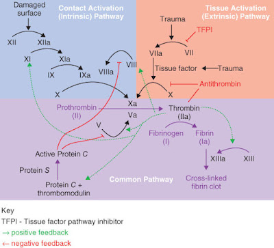
COAGULATION: FIBRINOGEN, PT, BT, TT, APTT.

FIBRINOGEN

Fibrinogen/Coagulation Factor I is a plasma glycoprotein produced by the liver. Fibrinogen is the action substrate for both thrombin (final coagulation enzyme) and plasmin (fibrinolytic system enzyme). Fibrinogen belongs to the acute phase proteins (appearing at 24-48 hours post-event). 

Defective fibrinogen levels show liver disfunction. Fibrinogen levels help assessing the body's ability to form a blood clot.  

While coagulation approaches its end, soluble fibrinogen is transformed into insoluble fibrinogen fibres. Fibrinogen fibres protect the wound by intercalating; along with the platelets, they form a clotted blood barrier that blocks bleeding until healing. 

Fibrinogen analysis measures the soluble amount of Factor I (fibrinogen dissolved in the blood), before its conversion into insoluble fibrin.

Fibrinogen normal levels: 200-400 mg/dL (according to the laboratory reference values - reactive used etc.).
 
 
PROTHROMBINE TIME (PT; INR): 

Prothrombin time (PT) evaluates the activity of factors involved in the extrinsic and common coagulation pathway. Evaluates both the activity of vitamin K-dependent coagulation factors (except F IX), factor V and fibrinogen, as well as the function of liver protein synthesis, with diagnostic and therapeutic implications. 

In vitro, main route of blood coagulation initiation: the extrinsic system. This includes blood components and vascular elements, the initiation of coagulation occurring when the tissue factor (FT) is bound to F VIIa. 
The F VIIa - FT enzyme complex activates both F IX and F X. 
F Xa interacts with its cofactor F Va, forming the prothrombinase complex; the complex: enough to generate few thrombin amounts next to FT-expressing cells. 

In vitro, PT detects clot formation. This represents fibrin polymerization, resulting from thrombin action.
Being absent in normal plasma, the tissue factor (FT) must be provided from an external source. This is why the cascade of enzymatic reactions triggered in PT is known as the "extrinsic pathway".  

Prolonged PT indicates deficiency of coagulation factors (I, II, V, VII, X) or an inhibitor's presence. 

PT is the most commonly used test monitoring oral anticoagulant therapy. Results may be expressed as follows:
- as clotting time - in seconds; PT - time (s); normal: 11-13.5 s;
- as a percentage (%) of normal prothrombin activity; measurable range: 12.5-120%; AP - volume fraction (%)
- as prothrombin ratio (PR = PT patient in seconds / normal plasma PT in seconds); PR - prothrombin time ratio (1)
- as INR - International Normalized Ratio - (1); normal: 0.8-1.1.
INR = (PT patient / PT normal plasma) x ISI; ISI: international sensitivity index of used thromboplastin, calculated in report to reference thromboplastin for which ISI = 1. 

INR = 2.0-3.0 target of oral anticoagulant treatment for most clinical cases;
INR = 2.5-3.5 target in anticoagulant treatment for: recurrent deep vein thrombosis, recurrent systemic embolism, cardiac stent, mechanical heart valves.


BLEEDING TIME (BT/CT)

A test that investigates primary haemostasis, thus being an indicator of the vascular and platelet phases efficiencies. 

BT depends on the:
- platelets (function and number)
- adhesion plasma proteins
- vascular wall matrix integrity
Critical values:  >15 minutes. 

Bleeding Time is increased when platelet levels are low or when platelets are qualitatively abnormal. 


THROMBIN TIME (TT)

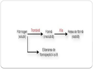
TT measures the time of fibrin formation under the action of thrombin, and its aggregation to form insoluble clot - the final coagulation step. Assesses the activity of fibrinogen.

Under thrombin action on fibrinogen, fibrinopeptides A and B are released. Upon their cleavage, fibrin monomers form soluble aggregates. 
Due to the action of Factor XIII (activated by thrombin), along with that of Ca ions, a transverse polymerization of fibrin monomers takes place, with the formation of insoluble fibrin. 


Thrombin Time mainly reflects the function and interaction between the exogenous thrombin and the endogenous fibrinogen. 

TT measurable range: 13-240 seconds. Thrombin clotting time is generally <22s, between 14-16s (every lab sets its own reference values, according to the reagent kits used; other labs may have: 7,0-12,0s). Critical values: >60s. 


ACTIVATED PARTIAL THROMBOPLASTINE TIME (APTT)

Also called Partial Thromboplastin Time (PTT), is a functional test evaluating both "intrinsic" and "common" coagulation pathways. 
In vitro, the contact system is involved when blood interacts with a foreign surface, such as the cardiopulmonary bypass. 

Kallikrein cleaves HK, releasing bradykinin and kinin-free kininogen (activated HK). 
In vitro, activation of HK is achieved by adding:
- "partial thromboplastin"; composed only of phospholipids; no protein, no TF;
- silica - a surface activator; provides the negatively charged surface.
Than, calcium chloride is added and time expressed in seconds is measured until the clot's formation. 
The so-called "partial thromboplastin" does not contain the tissue factor (protein) needed to initiate coagulation in PT, as in "complete thromboplastin" stage. The silica provides the negatively-charged surface needed for coagulation contact pathway activation. 

The cascade of reactions triggered in PTT were called as "intrinsic pathway" based on the misconception that coagulation is initiated without the addition of any external factors. The external factor involved in initiating coagulation in PTT: the negatively-charged glass surface of the reaction tube; this can be potentiated by adding silica, kaolin, ellagic acid, in what we now refer to as the "activated" partial thromboplastin test.  

Deficiency/inhibition of HK, prekalikrein or Factors XII, XI, IX and VIII causes prolongation of aPTT with normal PT, while deficiencies of "common pathway" coagulation factors (X, V, II, Fibrinogen) can prolong both aPTT and PT. aPTT is not influenced by FVII or FXIII deficiencies. 

Normal aPTT: 21-35s (therapeutic: 2,0-2,5x normal)


COAGULATION: 


"INTRINSIC PATHWAY": prekalikrein, high-molecular weight kininogen-HK, Factors XII, XI, IX and VIII;
"COMMON PATHWAY": Factors X, V, II and I. 


CLINICAL BLOOD HAEMOSTASIS - BLOOD TYPE IDENTIFICATION

 BLOOD TYPE IDENTIFICATION: Based on characterization of an individual's blood according to the presence/absence of an antigen on the surface of his erythrocytes. Most blood group antigens are glycoprotein in nature and genetically stable. 

The antigen-antibody reaction in which the blood group antigens and their specific antibodies participate is one of agglutination (of red blood cells). Hence, the antigens are also called agglutinogens and the antibodies also agglutinins.  

In current medical practice, the ABO and Rh systems are important. 

The main blood groups: 

A

B

AB

0.

Rh factor (Rhesus) is a group of antigens that may be present or absent on the surface of erythrocytes. Most people have Rh factor present on erythrocytes, so they are called Rh positive. Rh negative: do not have the Rh factor present. 

Incompatibility of Rh positive blood with Rh negative blood is an important cause of transfusion reactions and haemolytic diseases in newborns. 


                                BLOOD TYPE COMPATIBILITY RESUME

                                            +   : Rh present;     -   : Rh absent


MICROORGANISMS IDENTIFICATION - culture growth etc

Microscopy: particularly useful for bacteria identification.

Blood analysis (serology, blood culture etc): identification of microorganisms in blood.

Pharyngeal/nasal exudate, sputum, ocular/otic secretions: collection of biological samples for microbiological examination. 

Urinalysis: first morning urine, from the intermediate jet, after local toilet. Important tool for microorganism presence identification.

Coproparasitological examination, coproculture, from faeces.

Vaginal, urethral, vulvo-vaginal secretion examinations (exudates etc)

Various smears with specific colourings - microscopical examinations of morphology.  

Various molecular genetics techniques for virus identification: RT-PCR, ELISA, Western-Blot, immunological techniques (immunochromatography, immunodifusion, immunofluorescence) etc. 


LABORATORY DIAGNOSIS FOR VIRUSES:


- virus isolation on cell cultures

- viral genome highlighting by gene amplification reaction after reverse transcription (Real-Time Polymerase Chain Reaction - RT-PCR) - frequently used due to high precision

- serological identification - immunochromatography techniques (rapid tests), ELISA, Western Blot etc.


CULTIVATION OF VIRUSES


Viruses have obligatory intracellular parasitism, hence they do not grow on artificial environment/mediums. They can only be cultivated on live substrates:

- laboratory animals - limited use nowadays due to spreading of cell culture usage popularity;

- embryonated chicken egg - offer embryonic tissue for viruses culturing; important in vaccine preparation;

- cell cultures - the mostly used virus-host system in virology research. 

Example: Poliomyelitis

                 - indicated collection of pharyngeal exudate, blood, faeces, cerebrospinal fluid;

                 - isolation of virus on cell cultures; RT-PCR.


CULTIVATION OF BACTERIA ON MEDIA CULTURE:


Microscopical examination: first step to bacteria identification. Main elements to establish bacteria identity: 

- cilia

- capsule

- endospores. 


Morphological examination: on microscopic preparations, fixed and coloured: smears

Smears: obtained by spreading a bacterial colony on a clean and degreased blade, which will be dried, fixed and coloured, for examination under a microscope. 


Culture media - provides nutrients and physico-chemical conditions for bacteria growth;

Sowing - contact of pathological product with the culture medium;

Isolation - single colony transition to other culture medium => pure culture.


MEDIUMS: 


Physical classification:

- solid - reflecting colour, diameter, appearance, media adherence etc;

- liquid - turbidity and turbidity type, surface/bottom film/deposit formation.  

Examples: homogeneous turbidity - S. aureus; surface film - Vibrio cholerae, bottom deposit - Streptococcus pyogenes. 


In terms of composition, media can be:

- simple - simple agar;

- composed - besides the basics, also organic substances (serum, blood) required for special growth care; example: blood agar;

- special mediums - isolation, enrichment, differential. 


BACTERIA CLASSIFICATION:


1. Tinctorial affinity to Gram staining (most important staining, dividing bacteria in Gram-positives - lilac and Gram-negatives - red): 

a) Gram-positives: Streptococcus pneumoniae, Staphylococcus aureus;

b) Gram-negatives: Neisseria gonorrhoeae and meningitidis, Escherichia coli, Helicobacter pylori, Haemophilus influenzae. The Enterobacteriacee family - large number of Gram-negative bacillus species living in the intestines; the most important genre it includes are: Escherichia, Shigella, Salmonella, Klebsiella, Proteus. Enterobacteria classification according to the fermentation capacity of lactose: 

- Pathogenic Enterobacteria: lactose-negative: Salmonella, Shigella;

- Conditioned-pathogenic or non-pathogenic enterobacteria: 

                            a) lactose-positive: Escherichia, Klebsiella

                            b) lactose-negative: Proteus.


2. Need for oxygen:

a) aerobic bacteria: Staphylococcus aureus;

b) anaerobic bacteria: Clostridium botulinum.


3. Glucose fermentation capacity:

a) glucose-fermentative bacteria: Vibrio cholerae;

b) glucose-nonfermentative bacteria: Pseudomonas aeruginosa. 


4. Pathogenicity classification: 

a) nonpathogen bacteria: suboptimal conditions inside human host

b) pathogenic bacteria: always causing illness; example: Treponema pallidum. 

c) conditionate pathogenic bacteria: from normal flora, causing illness in various conditions.  


EXAMPLES OF AGARS AND COLORATIONS FREQUENTLY USED IN MICROBIOLOGY FOR BACTERIA IDENTIFICATION:

- MacConkey Agar: selective and differential culture medium for bacteria. Isolates Gram-negative and enteric bacteria. Differentiates them based on lactose fermentation. Lactose fermenters turn red or pink, non-fermenters do not change colour.

- Kligler's Iron Agar: Enterobacteriaceae identification, based on double sugar fermentation and hydrogen sulphide production.

- Ziehl-Neelsen stain: acid-fast stain identifying acid-fast organisms, mainly Mycobacteria (Nocardia as well). Acid-fast bacilli are bright red after staining. 

- Voges-Proskauer test: detects acetoin in a bacterial broth culture. Red: positive, yellow-brown: negative. Principle: digestion of glucose to acetylmethylcarbinol. 

- BD CLED Agar: CLED: Cystine-Lactose-Electrolyte-Deficient; differential culture medium for bacteria in urine; supports pathogen growth but prevents Proteus species accumulation due to the lack of electrolytes. 


DIAGNOSIS OF PARASITES:


- Coproparasitology: - faeces examination for parasites;

- ELISA;

- Direct agglutinations to detect IgM antibodies;

- Blood culture; cerebrospinal fluid examination for parasites;

- Smear and thick drop examination of peripheral blood; May-Grumwald-Giemsa staining;

- Immunological examination: ELISA, hemagglutination; latex agglutinations

- Parasites/parasite eggs identification in faeces and other material through microscopical examination.

- Macroscopical examination of faeces for parasite and/or parasite eggs elimination;


MICOLOGICAL DIAGNOSIS:


- based on the clinical lesion's aspect and microscopic analysis results of hairs and squamous material collected from lesions. 


ANTIBIOGRAM - drug choosing

 An antibiogram is an overall profile of antimicrobial susceptibility testing results of a specific microorganisms to a battery of antimicrobial drugs. 

It shows susceptibility of the microorganism investigated to particular antimicrobial drugs. Obtained results classified as: resistant, intermediate or sensitive to the action of a certain antibiotic. 


White disks: the antimicrobial drugs. Unaffected area around the white disks: growth of the microorganism i.e. the drug does not kill the microbe. Affected area around the disks (radii of inhibited areas) gets bigger (no organism growth) with as much as the drug can affect the microbe's growth. In clinics, this affected area is measured (to be taken into consideration for treatment choosing by the clinican), along with the name of each drug with annotations such as "microbe not resistant to drug X - diameter of inhibition zone measured: MIC (Y mm)".
X: name of the drug/antibiotic
Y: number measured in mm


MICROBIOLOGY - fast kits

VARIOUS RAPID MICROBIOLOGICAL EXAMINATIONS

    - include several rapid test kits with multimicrobial identification;

    - can be performed on blood, serum, urine or faecal material.

Example: 


MOST RECENT HEALTH NEWS!

Current news about health from all around the world


THE CLINICAL ANALYSIS LABORATORY - BASICS

BASICAL CLINICAL ANALYSIS LABORATORY


CLINICAL SERUM BIOCHEMISTRY


GLYCATED HEMOGLOBIN (BLOOD)


PROTEIN ELECTROFORESIS (SERUM)


URIANALYSIS (1st in the morning/8h URINE):

                     - BIOCHEMISTRY  OF URINE

                     - BASIC MICROSCOPY OF URINE (SEDIMENTATION)


CLINICAL IMMUNOLOGY (BLOOD AND FAECAL SAMPLES)

                    - AUTOMATIC

                    - RAPID MANUAL TESTS


IONOMETRY (SERUM/URINE): 

                            - SODIUM

                            - POTASSIUM

                            - OTHERS


CLINICAL BLOOD HAEMATOLOGY AND HAEMOSTASIS

            - HAEMATOLOGY

                    - COMPLETE BLOOD COUNT (CBC): RBCs, WBCs, PLTs

                    - BLOOD SEDIMENTATION (ESR-ERYTHROCYTE SEDIMENTATION RATE)

            - HAEMOSTASIS:

                    COAGULATION (PLASMA): FIBRINOGEN; PT, INR; CT/BT; TT; APTT                

                    - BLOOD TYPE IDENTIFICATION: A, B, O; Rh Factor


MICROBIOLOGY (SERUM/BLOOD, URINE, FAECAL, CEREBROSPINAL FLUID ETC): 

                     - MICROORGANISMS IDENTIFICATION

                                                             - CULTURE GROWTH

                                                             - ANTIBIOGRAM

                                                             - VARIOUS RAPID MICROBIOLOGICAL EXAMINATIONS