Search This Blog

Protein S-100

Role of S100 proteins in health and disease

- Assessment of S100 protein structure, function, and expression.
- Mechanism of action of S100 proteins in disease pathophysiology.
- S100 proteins as biomarkers for disease detection and prognosis.
- Therapeutic strategies targeting S100 proteins to treat disease. 


Introduction

The S100 family of proteins contains 25 known members that share a high degree of sequence and structural similarity. S100 family members are multifunctional proteins that regulate a diverse array of cellular processes including proliferation, differentiation, inflammation, migration and/or invasion, apoptosis, Ca2+ homeostasis, and energy metabolism. The S100 protein family is a multigene calcium-binding family, each encoded by a separate gene. S100 proteins exert their actions usually through calcium binding, although Zn2+ and Cu2+ have also been shown to regulate their biological activity. The S100 protein family is an acidic calcium-modulated protein family of low molecular weight (10-12 kDa), mainly expressed in vertebrates. S100 proteins have the ability to form homodimers, heterodimers and oligomers. Only a limited number of family members have been characterized in depth, and the roles of other members are likely undervalued. 
Biological functions of S100 proteins: a broad range of intracellular and extracellular functions. Intracellular functions include: regulation of enzyme activity and protein phosphorylation, calcium homeostasis, regulation of cytoskeletal components and of transcriptional factors. Extracellular functions of S100 proteins: act as Ca2+ sensor proteins in the cell and transmit a signal by Ca2+-dependent binding to the target protein, regulating its biological activity. Extracellularly in the presence of high concentrations of Ca2+ and Zn2+, S100 proteins can form polymers and bind to the receptor named RAGE. 
S100 protein reactivity: seen in 50-90% of tumors with extent and intensity of reactivity dependent on grade of tumor.

Due to the diverse range of cellular functions undertaken by S100 proteins, some family members have been given more than one name. These include: 
- S100A4 - calvasculin
- S100A6 - calcyclin
- the dimer formed by S100A8/A9 - calprotectin
- additionally, calgranulins comprise a group of S100 proteins including: 
                                         - S100A8 (calgranulin A)
                                         - S100A9 (calgranulin B)
                                         - S100A12 (calgranulin C)
                                            which act as sensors of intracellular Ca2+ levels 


S100 protein structure, expression, and function 

Molecular structure 
The S100s constitute a family of proteins where each protein is encoded by an individual gene. Of the 25 human S100 genes, 19 (group A S100 proteins) are located within chromosome 1q21. Other members (S100A11P, S100B, S100G, S100P and S100Z) map to different regions. Each member of the S100 protein family has a similar molecular mass of 10-12 KDa, and they each share 25-65% similarity in their amino acid sequence. Upon Ca2+ binding, the S100 proteins experience a conformational change that allows interaction with target proteins. The distribution of hydrophobic and charged residues, together with differences in surface configurations, contribute to the specific target binding patterns described amongst S100 family members.

Expression
Members of the S100 gene family show different patterns of both cell- and tissue-specific expression. Expression of S100 proteins is carefully regulated in order to ensure the maintenance of immune homeostasis. Calprotectin (S100A8/A9): constitutively expressed in certain immune cells (monocytes, neutrophils, dendritic cells). Upon activation, it is also expressed in fibroblasts or mature macrophages, amongst others. Epigenetic mechanisms play a vital role in S100 gene expression regulation (methylation of DNA CpG islands: a common method of transcriptional repression). DNA hypomethylation significantly induce expression of S100 members in prostate and gastric cancer.

Function
S100 proteins have been implicated in the control of a wide number of intracellular and/or extracellular functions, including regulation of cell apoptosis, proliferation, differentiation, migration/invasion, energy metabolism, Ca2+ homeostasis, protein phosphorylation and inflammation in different cell types.  

S100s as damage associated molecular pattern (DAMP) molecules
DAMPs play a key role in the pathogenesis of many inflammatory diseases, including rheumatoid arthritis, osteoarthritis and atherosclerosis. After cell damage/stress or activation of immune cells including neutrophils and macrophages, S100 proteins are released to the extracellular space where they play a key role in the regulation of several immune and inflammatory processes. They act as DAMP molecules to activate both immune and endothelial cells by binding to toll-like receptors (TLR)s and receptors for advanced-glycation end products (RAGE).

S100s in immune cell migration, invasion and differentiation
Increasing evidence shows that several S100 proteins contribute to leukocyte migration. For instance, as well as inducing pro-inflammatory cytokine production in macrophages through the activation of the NF-kB and p38 mitogen activated protein kinase (MAPK) pathways, S100A8/A9 has been seen to mediate immune cell migration; S100A12 has been shown to induce the production of pro-inflammatory cytokines interleukin (IL)-6 and -8 through RAGE-dependent NF-kB activation, resulting in the recruitment of monocytes; S100A10 has been reported to recruit macrophages to tumour sites; whereas S100A8/S100A9 have been shown to signal through RAGE to mediate the effect of TNF-a on the differentiation of myeloid-derived suppressor cells. 

S100s as biomarkers for disease
Since a number of S100 proteins can be identified in body fluids, they may be used as biomarkers to detect a specific disease, where their increased expression levels are indicative of pathological conditions. 
As such, S100A4 has been reported as a novel biomarker and an important regulator of glioma stem cells, with its increased expression contributing to the appearance of a metastatic phenotype, as well as having been described as a marker for lupus nephritis activity, a determinant factor for the onset of the complex inflammatory autoimmune disease lupus erythematosus; increased serum levels of S100A6 have been reported in patients with gastric cancer; S100A7 levels have been found to be increased in cerebrospinal fluid and brain of patients with Alzheimer's disease; blood levels of S100A12 are increased in patients with diabetes and it has also been used as a biomarker for detection of other inflammatory diseases such as systemic-onset juvenile idiopathic arthritis; augmented serum levels of 
S100A8/A9 have been seen in individuals with obesity and in patients with coronary artery diseases. Fig. 3
Importantly, S100A8/A9 has also proven to be a useful biomarker for disease activity in the management of inflammatory bowel diseases such as Crohn's disease, and faecal S100A8/A9 detection can ben used to differentiate inflammatory bowel disease from irritable bowel syndrome. 
Finally, evidence showed that S100B can be used as a monitoring and prediction tool for management of traumatic brain injury, while its overexpression has also been associated with certain genetic disorders such as Down syndrome and even to certain mood disorders as a consequence of glial pathology.

S100s as therapeutic targets 
S100 proteins, particularly calgranulins (S100A8, A9 and A12), play a major role in the mediation of the immune responses characteristic of a series of diseases, including inflammatory arthritis, atherosclerosis and microbial infections, as well as joint inflammation and cartilage degradation in patients with rheumatoid arthritis. S100A7 has been found to be abundantly expressed in psoriatic lesions or in serum from psoriatic patients as well as in dermatitis skin lesions, induced by pro-inflammatory factors such as TNF-a, IL-17 and IL-22.
Several S100 proteins bind to TLR4 and RAGE. Importantly, the heterodimeric form of S100A8/S100A9 can bind TLR4, whereas high extracellular Ca2+ concentrations induce the formation of S100A8/S100A9 tetramers, preventing its interaction with TLR4, thus providing an autoinhibitory mechanism for modulating S100A8/9 biological activity.
The use of S100 function-blocking antibodies might provide an effective therapeutic strategy to treat cancers and immune disorders. Anti-allergic drugs have been reported to bind to S100A12, blocking downstream RAGE signalling and subsequent NF-kB activation.
Extracellular S100A8/S100A9 levels: closely linked to inflammatory and autoimmune diseases (rheumatoid arthritis, inflammatory bowel disease, cystic fibrosis, diabetic nephropathy, cardiovascular disease). S100A8 would be a good target against obesity-induced chronic inflammation. Increased S100A8/A9 expression in the tumour microenvironment is associated with the progression and aggressiveness of the disease.

S100A4
S100A4 plays a significant role in many physiological functions including cell motility, adhesion, proliferation, invasion, and metastasis. Intracellular S100A4 binds to proteins of the cytoskeleton including F-actin and non-muscle myosin heavy chains, both involved in cellular stability and/or motility. By contrast, extracellular S100A4 regulates the expression of extracellular matrix (ECM)-remodelling enzymes such as MMPs, which are implicated in mediating cellular migration in various tissues, and can signal through membrane receptors to activate proinflammatory pathways.  
Fig. 2

S100A4 in disease
S100A4 and cancer
S100A4, together with many other proteins: involved in the complex multi-step process of cancer metastasis at the molecular level. S100A4, secreted from both tumour and non-malignant cells, plays a key role in the regulation of angiogenesis, cell migration and inflammation.
S100A4 levels are increased in many types of cancers and tumour microenvironments, including brain, breast, lung, gastric, liver, pancreatic, colorectal and prostate cancers amongst others, in addition to osteosarcoma, leukaemia and malignant melanoma, always associated to poor prognosis. S100A4 is a strong likely biomarker for cancer diagnosis and metastasis prediction.
S100A4 and non-cancer pathologies
Even though S100A4 is best known in a disease context for its participation in cancer progression and metastasis, an increase in S100A4 expression has also been associated with several non-tumour pathophysiological processes including tissue fibrosis, inflammation, neuroprotection and cardiovascular events.  

S100 family signaling network and related proteins in pancreatic cancer
Multiple proteins of the S100 protein family are closely related to pancreatic cancer, including the following proteins: S100A2, S100A4, S100A6, S100P, S100A11.
S100 proteins interact with receptor for advanced glycation end-products (RAGE), p53 and p21, which play a role in the degradation of the extracellular matrix (ECM) and metastasis, and also interact with cytoskeletal proteins and the plasma membrane in pancreatic cancer progression and metastasis. S100A11 and S100P are significant tumour markers for pancreatic cancer and unfavorable predictors for the prognosis of patients who have undergone surgical resection. S100A2 has been suggested to be a negative prognosis biomarker in pancreatic cancer, and the expression of S100A6 may be an independent-prognosis impact factor. The expression of S100A4 and S100P is associated with drug resistance and differentiation, metastasis and clinical outcome. Conclusion: S100 proteins may be used as molecular markers for the early diagnosis, treatment and prognosis of pancreatic cancer.  

The S100 family: intracellular and extracellular activities
Intracellular activities of S100 proteins 
Members of the S100 family interact with p53 and this produces differential effects, depending on the activity of the protein involved. Both S100A4 and S100B are thought to inhibit p53 phosphorylation, leading to the inhibition of this transcriptional activity, thereby compromising p53 tumor-suppressor activity. By contrast, S100A2 promotes p53 transcriptional activity. Thus the balanced actions of different S100 proteins within a cell determine its function. Many of the S100 family members are involved in modulating cytoskeletal dynamics.      
Extracellular roles of S100 proteins
S100 proteins are involved in the extracellular stimulation of neuronal survival, differentiation and astrocyte proliferation, resulting in neuronal death via apoptosis, and stimulate (in some cases) or inhibit (in other cases) the activity of inflammatory cells. S100 proteins are closely related to a variety of human diseases, such as neurological disorders, cancer, inflammation and heart disease.


S100 proteins: methods of measurement
Analytical methods such as immunoradiometric assay (IRMA), mass spectroscopy, western blot, ELISA (enzyme linked immunosorbent assay), electrochemiluminence and quantitative PCR, can detect S100 changes in immunohistochemical expression or in serum concentration with high sensitivity, providing an important tool in clinical diagnosis.


S100 expression in related diseases
Diseases associated with altered expression levels of S100 proteins can be classified into four categories:

1. Neurologic disorders
As protein S100B is primarily produced by astrocytes in CNS, its increased expression - as well as that of glial fibrillary acidic protein (GFAP) - represents a hallmark of astrocytic activation. S100B protein's autocrine effects on astrocytes (upregulation of IL-6, TNF-alpha expression) are mediated through its interaction with RAGE (Receptor for Advanced Glycation End products). Secretion of S100B is an early process during the glial response to metabolic injury (oxygen, serum and glucose deprivation). 
Elevated levels are observed in patients suffering from chronic neurodegenerative disorders such as Alzheimer's disease. Elevated levels of S100B originating from necrotic tissues might enhance or even amplify neurodegeneration by S100B-induced apoptosis.
It has been reported that serum and CSF S100 level is raised in systemic lupus erythematous with neuropsychiatric involvement (i.e. organic brain syndrome, seizures, cerebral vascular accident, psychosis) and in obstructive sleep apnea syndrome, reflecting the ongoing neurological damage. Significant increase of serum S100B is observed during exacerbations of bipolar disorder (episodes of mania and depression).
The above described wide range of applications has led to the consideration of S100B measurement in neurologic disorders as analogous to that of CRP in systemic inflammation.

2. Neoplastic disorders
Different forms of cancer exhibit dramatic changes in the expression of S100 proteins such as S100B, S100A2, S100A4, S100A6, and S100P. The S100-RAGE signalling pathway plays an important role in linking inflammation and cancer and in tumour cell survival and malignant progression (RAGE-deficient tumours are characterized by accelerated apoptosis, reduced activation of NFkB and significantly impaired proliferation).
Elevated levels of S100A4 (metastasin) are associated with poor survival rates in breast cancer patients. Increased serum concentration of S100A4 is also found in esophageal squamous and colon carcinoma, invasive pancreatic carcinoma, non small cell lung cancer, bladder carcinoma and correlates with a worse outcome and more aggressive disease. 
Concerning the detection of brain metastases, serum concentration of S100B has a good negative predictive value. As its levels may also reflect the existence of cerebrovascular ischemic changes without infiltrating tumour, it may be used in conjunction with proApolipoprotein A1 for a sufficiently specific serum-based diagnostic of the presence of metastatic brain tumours.
S100A2 is highly expressed in tumours such as: non-small lung cancer, gastric/oesophagea squamos cancer, lymphoma, granular cell tumours of the gastrointestinal tract, renal tumours, papillary and anaplastic thyroid carcinomas. 
There is accumulating evidence that BRCA1 negative breast carcinomas exhibit increased expression of S100A7.
S100A8 and S100A9 form a heterodimer complex implicated in regulating cell proliferation and in the metastatic process.

Summary of the links that have been noted between various types of cancer and members of the S100 protein family:
Cancer                           Members of the S100 protein family
Melanoma                     S100B (established use), S100A4, S100A2
Breast                            S100A4, S100A7 (promising results)
                                      S100A8, S100A9, S100A2, S100A6, S100A11
Pancreatic                     S100A4, S100A10, S100A11, S100P (8-fold increase)
Colorectal                     S100A4, S100A6, S100A8, S100A9, S100A11
Gastric                          S100A2, S100A4, S100A8, S100A9, S100A11
Bladder                         S100A4, S100A11 (down-regulation associated with decreased survival)
Ovarian                         S100A1, S100A4
Prostate                         S100A2, S100A4, S100A11 (up-regulation associated with advanced stage)
Lung (squamous cell)   S100A2, S100A4, S100P
Renal                             S100A1, S100A11,
                                      S100A2 (3.8-fold decrease in 93% of patients)
Thyroid                         S100A2, S100A4
Lymphoma                    S100A2, S100P

3. Cardiac diseases
S100A1 is specifically and highly expressed in the mammalian myocardium, where it modulates contractile performance of the heart via interaction with contractile filaments and with proteins of the sarcoplasmic reticulum (SR). It also increases the release of calcium from the sarcoplasmic reticulum by interacting with the ryanodine receptor. S100A1 is up-regulated in right ventricular hypertrophy and down-regulated in end-stage heart failure, indicating a correlation between S100A1 expression and contractile performance. 

4. Inflammatory diseases
S100A8, S100A9, and S100A12, are predominantly expressed in phagocytes and are strongly associated with pro-inflammatory functions. They are secreted especially at sites of inflammation. The serum concentrations of these S100 proteins correlate with inflammatory disease activity; high levels were identified in several inflammatory disorders such as rheumatoid arthritis, chronic bronchitis, and cystic fibrosis. S100A7, S100A8, S100A9, and S100A12 are up-regulated in active psoriatic lesions. Overexpression of S100A7 (acting as a keratinocyte- derived chemotactic agent for immune cells) is also seen in many epidermal inflammatory diseases, like atopic dermatitis, mycosis fungoides and Darier's disease. Antiallergic drugs which bind to S100A12 might block the S100 protein-RAGE interaction, implying a promising approach to anti-inflammatory therapy. Enteric glial-derived S100B (associated with the onset of inflammation) is increased in the duodenum of patients with celiac disease and contributes to nitric oxide production. 


Conclusions

Characterization of primary tumor lesions has been used to identify and evaluate the risk in the development of tumor metastasis and to predict prognosis and therapy responses in various types of cancer. As a result, several S100 members, mainly S100A4 and S100A8/9, have been identified as key players in the pathogenesis of many types of cancer, as well as of several other disease conditions including diabetes and other inflammatory diseases. Elucidating the mechanisms of action of S100 proteins in the pathophysiology of these diseases may therefore lead to the development and application of novel, more effective therapeutic approaches. S100 proteins can be used as biomarkers in early disease detection and prognosis, and in the development of novel strategies based around anti-S100 therapies.

The members of the S100 protein family, through their interaction with several effector proteins, are involved in the regulation of a diverse spectrum of cellular processes. Although their pathophysiologic implications still require further clarification, some of these proteins have already been successfully investigated in clinical context.

Members of the S100 protein family proved to be useful biomarkers in clinical applications and the S100 protein-targeted therapies emerge as useful opportunities in specific clinical settings.  

PTKs: Protein Tyrosine Kinases

    Protein tyrosine kinases (PTKs) are a group of enzymes that play crucial roles in cellular communication and regulation. PTKs control key functions of normal and malignant cells. They are ubiquitous enzymes that are integrally involved in the regulation of transformation mechanisms, normal and pathological growth, immune responses, and a variety of intracellular signaling mechanisms. 


PTKs play a fundamental role in various aspects of cell biology, including cell proliferation, survival, adhesion and motility by regulating ligand-mediated signal transduction, cell cycle regulation and progression, cell division, cell differentiation and cytoskeleton functions. Hence, the importance of PTKs in cell growth regulation on multiple levels is evident. Tyrosine kinases have major clinical implications: they activate lymphocytes and mediate communication in cell types like adrenal chromaffin cells, platelets, and neural cells. Dysregulation of PTK-activated pathways, due to mutations, often by receptor overexpression, gene amplification, or genetic mutation, is a causal factor underlying numerous cancers (initiation and progression). Many of the PTKs represent proto-oncogenes and their mutations and/or abnormal expression result in the acquisition of malignant phenotype by the affected cells. In all likelihood, specific types of lymphomas as well as other types of malignancies display distinct patterns of expression of tyrosine kinases (besides other gene families). Identification of such patterns may play an important role in diagnosis of malignant tumors, particularly if the conventional methods yield equivocal results.

    PTKs are some of the most frequently altered genes in cancer, either via mutation, overexpression, or amplification. The resultant deregulated cellular signaling contributes to disease progression and drug resistance. Regulation of PTKs is controlled both by extensive post-translational modifications, particularly protein phosphorylation and by changes in PTK abundance. Thus, there is utility in quantifying the expression of PTKs to identify drug response signatures and reveal new biological characteristics.

    A tyrosine kinase is an enzyme that transfers a phosphate group from ATP to specific tyrosine residues within proteins. This process acts as an "on" or "off" switch in various cellular functions. PTKs hence mediate the enzymatic transfer of the gamma phosphate of ATP to the phenolic groups on tyrosine residues to generate phosphate monoesters.  

- Tyrosine kinases belong to the broader class of protein kinases, which also phosphorylate other amino acids like serine and threonine.  

- Phosphorylation by kinases is essential for signal transduction within cells, regulating activities such as cell division. 

- These enzymes participate in extracellular signal transmission, affecting gene expression in the nucleus. 

- Mutations in tyrosine kinases can lead to uncontrolled cell growth, a hallmark of cancer. Kinase inhibitors are effective cancer treatments. 

    PTK family of enzymes is generally divided into two groups: receptor PTKs (with more than twelve distinct families) and nonreceptor PTKs (with more than nine distinct families). 
    Receptor and Non-Receptor Tyrosine Kinases
    - Receptor Tyrosine Kinases (RTKs): These function in transmembrane signaling, receiving signals from outside the cell; 
     - Non-Receptor Tyrosine Kinases: Operate within the cell, participating in signal transduction to the nucleus.    

    Typically, expression of PTKs is measured by enzyme-linked immunosorbent assay, fluorescence activated cell sorting and immunoblotting, which provide information for a limited number of proteins in a single assay. Multiplexed targeted proteomic assays, on the other hand, could reveal simultaneous alterations of protein expression in entire PTK pathways. A widely used targeted proteomics approach for quantification is multiple reaction monitoring (MRM, also termed selected reaction monitoring), technique that has both advantages and disadvantages. 
    In summary, protein tyrosine kinases are pivotal players in cellular communication, and understanding their roles helps uncover new therapeutic strategies. Many PTKs have been shown to act as oncogenes and analysis of PTK expression by malignant cells will lead to a better understanding of oncogenesis. This in turn will lead to novel therapies based on selective inhibition of PTKs. The approach has already been proven effective in chronic myeloid leukemia and related bcr/abl-PTK-positive disorders. Expression of PTKs may be determined by RT-PCR using degenerate primers which recognize common, relatively invariable cDNA sequences of members of the PTK family. Other approaches to determine PTKs are also available nowadays, as it was previously mentioned. 

    PTKs are among the most intensively pursued superfamilies of enzymes as targets for anti-cancer drugs. PTK expression varies between different types and stages of cancer and alterations in PTK expression are an important mechanism of resistance to targeted cancer therapeutics. These considerations suggest that multiplexed, targeted analysis of PTK expression profiles are valuable in studying mechanisms of drug susceptibility and resistance. PTK profiling at the protein expression level may provide a robust alternative to study adaptation of signaling networks in human tumors.   

RAPID TESTS: AGGLUTINATION AND IMMUNOLOGY TESTING

RAPID AGGLUTINATION LATEX TESTS - from blood:

- ASLO;

- CRP;

- RF.


HEPATITIS: A, B, C, D and E:

- HEPATITIS A VIRUS - from blood;

- HEPATITIS B VIRUS - from blood ;       

- HEPATITIS C VIRUS - from blood;

- HEPATITIS D VIRUS - from blood;

- HEPATITIS E VIRUS - from blood, stool. 


SYPHILIS - treponema pallidum; from blood and other fluids:

- VDRL;

- RPR;

- TPHA.


HIV TESTING - from blood, oral fluid, urine.


TROPONIN LEVEL TESTING - from blood. 


HELICOBACTER PYLORI DETECTION: 

- ANTIBODY DETECTION - from blood;

- ANTIGEN TEST - from stool.


FAECAL OCCULT BLOOD (FOB) TEST - from stool.


GIARDIA LAMBLIA - ANTIGEN DETECTION - from stool.


IMMUNOGLOBULIN E - from blood.


CORONAVIRUS - from blood, nose/throat swab. 


ASLO, CRP, RF: rapid agglutination tests

Serology - Immunology

Agglutination Kits & Serology Reagents: there are many options of latex serology kits on the market. Common serological tests to detect in serum:
- ASLO/ASO: Anti-streptolysin-O;
- CRP: C-Reactive Protein;
- RF: Rheumatoid Factor.

The latex assays are available in different kit sizes, with positive and negative controls provided. The full kits include all the required consumables. They use the common agglutination method for efficient, reliable testing, to ascertain the causes of inflammation, rheumatoid disorders and streptococcal infections. Latex agglutination testing is sometimes referred to as latex serology. 
Agglutination occurs when an antigen comes into contact with its corresponding antibody. The rapid test kits are used to identify streptococcal infections, levels of C-reactive protein that would indicate high levels of inflammation in the body and rheumatoid factors that would indicate a diagnosis of rheumatoid arthritis.

Latex serology tests are used to ascertain whether specific antigens or antibodies are present in a sample. This is done by applying the samples to the latex beads. If the suspected substance is present in the sample, the latex beads clump together (agglutinate). The results of latex serology tests can be obtained within 15 minutes to an hour, allowing a rapid diagnosis to be made, which is obviously a significant advantage.
 

These slide agglutination assay tests are used for the qualitative and semi-quantitative determination of ASLO, CRP and RF in human serum. The simple, five-step procedure provides easy-to-read results in 2 minutes. No initial dilution of patient samples is required. Kits usually include ASLO/CRP/RF latex reagents, reactive control, nonreactive control, 100 disposable stirrer pipettes and disposable cards. The latex reagent does not require additional preparation. MATERIAL REQUIRED: automatic pipettes, mechanical rotator, adjustable at 100 r.p.m, laboratory alarm clock.

QUALITY CONTROL: Positive and negative controls should be run daily following the steps outlined in the Qualitative Test, in order to check the optimal reactivity of the reagent. The positive control should produce clear agglutination. If the expected result is not obtained, you must not use it. 

ASO/ASLO: slide agglutination test for the qualitative and semi-quantitative detection of streptolysin antibodies in human serum. Analytical sensitivity: 200 (+/-50) IU/mL. 
CRP: slide agglutination test for the qualitative and semi-quantitative detection of C-reactive protein in human serum. Analytical sensitivity: 6 mg/L. 
RF: slide agglutination test for the qualitative and semi-quantitative detection of rheumatoid factors in human serum. Analytical sensitivity: 8 IU/mL. 

SOURCES OF ERROR: Bacterial contamination of controls and specimens as well as freezing and thawing of the ASLO/CRP/RF-latex reagents may lead to false positive results. Traces of detergent in the test cards may give false positive results. Preferably the used cards would not be reused. If cards are reused, wash them first under tap water until all reactants are removed and then with distilled water. Allow to air dry, avoiding the use of organic solvents as they may impair the special finish on the slide. 
The ASLO/CRP/RF-latex antigen must not be used beyond its expiry date because a prolonged storage can affect the sensitivity of the suspension. Do not allow reagents in the kit to get in contact with skin or mucous membranes.


Reagents are stable if stored at 2-8ºC up to expiration date. Allow the suspension to reach to room temperature and mix gently prior to use. Do not freeze. Frozen reagents could change the functionality of the test. Reagent and Controls are ready for use and stable until the expiry date stated on the label. Do not use haemolysed, lipaemic or contaminated serum for testing. Hemoglobin (<10 g/L), bilirubin (<20 mg/dL) and lipemia (<10 g/L) do not interfere. Other substances (rare) may interfere. SAMPLES: must be fresh, clear serum. After the clear serum has been separated it may be stored at 2-8ºC for up to one week or longer periods at -20ºC, before testing. Undiluted samples should be used. Do not use plasma since fibrinogen can form non-specific agglutination. The sensitivity of the test may be reduced at low temperatures. The best results are achieved at 15-25ºC. 


PROCEDURAL STEPS for the QUALITATIVE TEST
1. Bring the test reagents and samples to room temperature. 
2. Transfer 50µl/1 drop of the patient's serum into one of the circles of the test card. Dispense 1 drop of positive control and 1 drop of negative control into two additional circles. 
3. Gently shake the suspension of the latex reagent (ASLO/CRP/RF), then using the pipette, aspirate dropper several times to obtain a thorough mixing. Add 1 drop/50µl reagent of the suspension to the same test circle with the patient's serum and to those with the controls. 
4. Using the stirrers, mix the serum and the latex reagent and spread them. Mix the contents of each circle while spreading over the entire area enclosed by the ring. Use separate stirrers for each mixture. 
5. Rotate the test card/slide for 2 minutes by means of a mechanical rotator (100r.p.m.) and observe immediately under a suitable light source for any degree of agglutination. 

READING THE LATEX SLIDES:

- Nonreactive: smooth suspension with no visible agglutination, as shown by the negative control. 
- Reactive: any degree of agglutination visible macroscopically (best to check under a suitable light). 

The delays in reading the results may generate in overestimation of the antibody present.


For the SEMI-QUANTITATIVE TEST:

- Dilute the sample to be tested following the 2-fold dilutions, as follows: 1/2, 1/4, 1/8, 1/16, 1/32. Follow the other steps as in the quantitative test.

How to make the dilutions: it is up to the biologist, for as long as it is correctly diluted mathematically. 

Example of dilution 1/2: 50µnon-diluted sample + 50µL dilution liquid (physiological serum/distilled water), then transfer 1 drop/50µL from the ASLO/CRP/RF latex reagent over the mixture. The other steps are as in the quantitative test.   

Reading is the same for both the Qualitative and for the Semi-Quantitative Test:
- Negative/Non-reactive: no agglutination; 
- Positive/Reactive: visible agglutination.  

The titer of the specimen for the Semi-Quantitative test is reported as the highest dilution that shows reactivity. The next higher dilution should be negative. If the highest dilution tested is reactive, repeat the test using the next dilution. 
The results obtained for the Semi-Quantitative Test do not replace the fully-automated quantitative tests available on the market. The levels obtained following the Semi-Quantitative Test procedure are considered only "approximate", not exact values, hence the naming of "Semi-Quantitative Test". 

The approximate ASLO/CRP/RF levels (IU/mL) present in the sample may be obtained by multiplying the titer of the last positive dilution by the minimum detectable unit (analytical sensitivity) corresponding to each reagent type. 


Reading the Semi-Quantitative tests:

ASLO: 
- 1/2 dilution shows reactivity => ASLO level: 2 x 200 = 400 IU/mL
- 1/4 dilution shows reactivity => ASLO level: 4 x 200 = 800 IU/mL
1/8 dilution shows reactivity => ASLO level: 8 x 200 = 1600 IU/mL
- 1/16 dilution shows reactivity => ASLO level: 16 x 200 = 3200 IU/mL

1/4 reactive and 1/8 non-reactive => ASLO level: 800-1600; ASLO>800 and <1600.

CRP: 
- 1/2 dilution shows reactivity => CRP level: 2 x 6 = 12 mg/L
- 1/4 dilution shows reactivity => CRP level: 4 x 6 = 24 mg/L
1/8 dilution shows reactivity => CRP level: 8 x 6 = 48 mg/L
- 1/16 dilution shows reactivity => CRP level: 16 x 6 = 96 mg/L

1/2 reactive and 1/4 non-reactive => CRP level: 12-24; CRP>12 and <24.

RF: 
- 1/2 dilution shows reactivity => RF level: 2 x 8 = 16 IU/mL
- 1/4 dilution shows reactivity => RF level: 4 x 8 = 32 IU/mL
1/8 dilution shows reactivity => RF level: 8 x 8 = 64 IU/mL
- 1/16 dilution shows reactivity => RF level: 16 x 8 = 128 IU/mL.

1/8 reactive and 1/16 non-reactive => RF level: 64-128; RF>64 and <128.

ASLO

Antistreptolysin O (ASO/ASLO)
 
- ASLO/ASO latex kit test: an indirect agglutination kit for the detection of anti-streptolysin-O in human serum using latex particles coated with streptolysing-O. 

ASO titer is adviced for the diagnosis of post infectious glomerulonephritis, scarlet fever, streptococcus infections and othersThe Streptolysin O is antigenic and cause hemolysis of RBCs. So there is antibody formation (Antistreptolysin-Ab) in the blood. 
Anti-Streptolysin O (ASO or ASLO) is the antibody made against streptolysin O, an immunogenic, oxygen-labile hemolytic toxin produced by most strains of group A and many strains of groups C and G streptococci. 
In the course of streptococcal infections, the extracellular products of the bacteria act as antigens to which the body responds by producing specific antibodies. Streptolysin O (ASLO) is one of two hemolysins (the other being Streptolysin S) produced by virtually all strains of Streptococcus pyogenes.  

Principle:
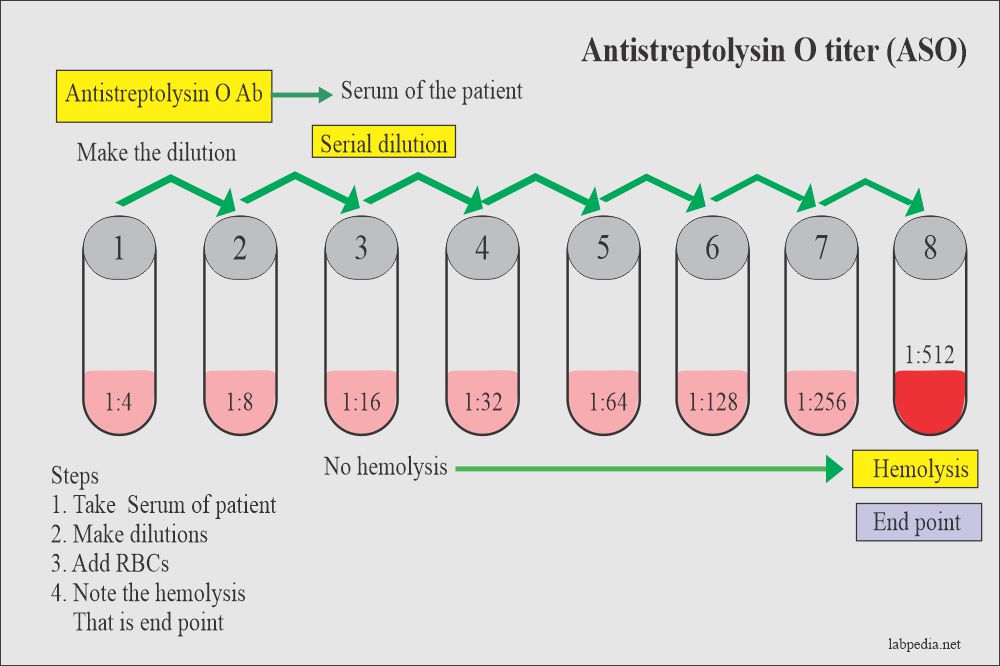
Patient serum with dilution is mixed with a fixed amount of streptolysin O- Ag and a reaction takes place. Then add Ab-coated RBC. There will be hemolysis in the tube, where streptolysin O is free.

Antistreptolysin (ASO) leading to hemolysis

The result is reciprocal of the highest dilution where hemolysis starts. Units are international units (IU).

Antistreptolysin (ASO) titer procedure

Normal value: below 200.
Definitive value: 400 or more.
Rising titer from 200-250 is significant.

ASLO-Latex Test: The assay is performed by testing a suspension of latex particles coated with streptolysin O antigen against unknown serum. The presence or absence of visible agglutination indicates the presence or absence of ASLO in the samples tested. 
REAGENT COMPOSITION ASLO-Latex Antigen. Suspension of polystyrene latex particles coated with stabilized streptolysin O in a buffered saline solution.
CONTROL + Human serum with an ASLO activity > 200 IU/mL.
CONTROL - Animal serum with an ASLO activity < 100 IU/mL. 


Precautions: Components of different human origin have been tested and found to be negative for the presence of antibodies anti-HIV 1+2 and anti-HCV, as well as for HBsAg.  Do not allow to contact with skin or mucous membranes.  

EXPECTED VALUES: 95% of healthy adults have ASLO titers of 200 IU/mL or less, the highest titers been found in school children with titers up to 250 IU/mL. Since a single ASLO determination does not provide much information unless it is high, titrations at bi-weekly intervals for 4 to 6 weeks of the doubtful cases are advisable to follow the evolution of the disease.
CLINICAL SIGNIFICANCE: Elevated ASLO serum titers occur in response to infection with hemolytic streptococci of Group A, C and G, producers of streptolysin O, an extracellular protein of enzymatic character with strong antigenic properties. Immunochemical assay of these specific antibodies to streptococcal metabolites provide valuable information to establish a diagnosis of streptococcal infections (acute rheumatic fever, glomerulonephritis). ASLO testing has a high diagnostic value on a tentative diagnosis made on the basis of case history and clinical findings. 
ANALYTICAL PERFORMANCE: The minimum detectable unit (analytical sensitivity) is of approximately 200 IU/mL (± 50 IU/mL). 
LIMITATIONS OF PROCEDURE: Positive reactions do occur in conditions other than glomerulonephritis, in which the production of ASLO is especially high. In scarlet fever, early and acute periods of rheumatoid arthritis, healthy carriers, complicated and no complicated tonsillitis and various streptococcal infections increased ASLO levels have been found. Biologically false negative reactions can occur in early infections and during the early years of life (from six months to 2 years). The sensitivity of the test may be reduced at low temperatures. The best results are achieved at 15-25ºC. Delays in reading the results may result in over-estimation of the antibody present. 

CRP

C-reactive protein (CRP)  

Definition
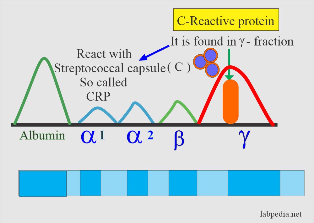
    1. CRP is γ-globulin (in the γ-region); is found in various inflammatory diseases. This is also called acute-phase protein. 
    2. CRP is produced in the liver.
    3. CRP name is derived from the reaction with streptococcal capsular (C) polysaccharide. 

C-Reactive Protein (CRP)                                                                C-Reactive Protein (CRP)

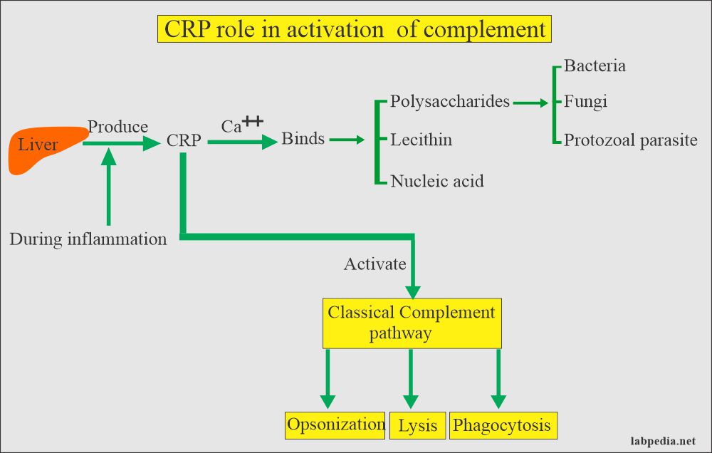
CLINICAL SIGNIFICANCE: C-reactive protein is an acute phase protein present in normal serum, which increases significantly after most forms of tissue injuries, bacterial and virus infections, inflammation, and malignant neoplasia. CRP contributes to non-specific defense by complement activation and accelerating phagocytosis. CRP testing has a high diagnostic value on a tentative diagnosis made on the basis of case history and clinical findings. 

Principle:
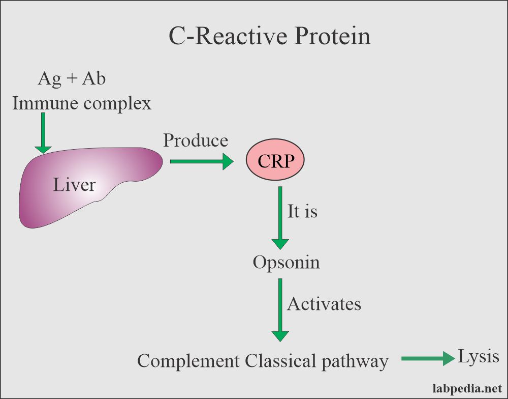
1. CRP is opsonin and it activates the complement system and ultimately leads to lysis.  

C-Reactive protein (CRP) leads to lysis by the activation of complement                         C-reactive protein (CRP) leads to lysis by the activation of complement 

C-Reactive protein (CRP) role in Complement activation                                      C-reactive protein (CRP) role in Complement activation

2. Serum of the patient (CRP) + Somatic C polysaccharide of pneumococci mixed and gives rise to a precipitate.
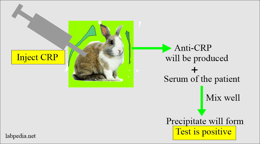
3. Practically CRP is injected into the rabbit when the anti-CRP antibody is produced. Now take serum of the patient (CRP) + Mix anti-CRP. This will give precipitation. 
                                                                              
CRP + Anti CRP = Precipitation

C-Reactive Protein (CRP) formationC-Reactive Protein (CRP) formation 

Causes of CRP
    1. Produced in various bacterial diseases.
    2. Produced by injured myocardial muscle in myocardial infarction.
    3. Positive in acute and chronic rheumatic fever.

CRP is also positive in:
    1. Sydenham's Chorea.
    2. Myocardial infarction and negative in angina.
    3. Many malignancies.
    4. Rheumatoid arthritis.
    5. Gout.
    6. Viral infection like Viral Hepatitis.
    7. Bacterial Pneumonia.
    8. Active Tuberculosis.
    9. Lepromatous Leprosy.
    10. Acute Tonsillitis, Scarlet fever, and Mumps.

PRINCIPLE: CRP-Latex Test is a rapid slide agglutination procedure, developed for the direct detection and semi-quantitation of C-reactive protein (CRP) in serum. The assay is performed by testing a suspension of latex particles coated with anti-human CRP antibodies against unknown serum. The presence of a visible agglutination indicates an increase of the CRP level above the upper limit of the reference interval in the samples tested. 
REAGENT COMPOSITION - CRP-Latex Reagent: Suspension of polystyrene latex particles coated with specific anti-human C-reactive protein antibodies in a buffered saline solution. CONTROL + Human serum with a CRP concentration > 15 mg/L. CONTROL - Animal serum with a maximum concentration of human CRP of 1 mg/L. Precautions: Components of different human origin have been tested and found to be negative for the presence of antibodies anti-HIV 1+2 and anti-HCV, as well as for HBsAg. However, the controls should be handled cautiously as potentially infectious. 
EXPECTED VALUES: While the C-reactive protein concentration is generally below 5 mg/L in the sera of healthy adults, in a number of disease states these values often exceeded within 4 to 8 hours after an acute event and reach levels up to 500 mg/L. Since an elevated CRP level is always associated with pathological changes, determination of CRP is of great value in diagnosis, treatment and monitoring of inflammatory conditions. 
ANALYTICAL PERFORMANCE The minimum detectable unit (analytical sensitivity) is of approximately 6 mg/L (5-10 mg/L). Hemoglobin (<10 g/L), bilirubin (<20 mg/dL) and lipemia (<10 g/L) do not interfere. Rheumatoid factors (>100 IU/mL) interfere. Other substances may interfere. 

LIMITATIONS OF PROCEDURE  The presence of rheumatoid factors (RF) in a serum sample may cause false positive reactions. The sensitivity of the test may be reduced at low temperatures. The best results are achieved at 15-25ºC. Delays in reading the results may result in over-estimation of the CRP concentration. In case of positivity, use the titration/dilution procedure, as presented above, at the "ASLO/CRP/RF" section.